Unit - 9
Microbiology
An organism who lives a life which is independent. Single cell organisms are organisms that have a single cell, they perform the entire activities with the single cell itself.
Single Celled Organisms
Protozoa: These are the Eukaryotic cells most of them live in water, either in fresh water or marine water very few are found in the soil or land. They are a variety of protozoa and some are parasites and some are free living. Some of them are known to even cause severe diseases in humans.
Their Internal structure generally consists of a nucleus, plasma membrane, cilia, mitochondria, vesicles, and other cell organelles. Though they are free living they are dependent on other sources for their food. They eat up bacteria and other organic matter.
Examples include: Amoeba, euglena, paramecium.
Parasites include: Entamoeba, plasmodium
Bacteria: These are prokaryotic single celled organisms. They are found to be present almost everywhere. They are found in soil, water, air and even in extreme climate conditions like heat and cold. There are many bacteria that have been affecting humans, causing a number of diseases since centuries. These bacteria have ability to obtain energy from various sources. Some of them are parasites, some autotrophs, saprophytes, and even chemotrophs.
Examples include E-coli, staphylococcus bacteria, salmonella typhi, pseudomonas aerogenosa etc.
These organisms have different ways to gain energy. Some cells absorb energy through the surface of the cell directly into the cell body. If the cell is large, the cell literally wraps its body around a small cell or can be a nutrient, absorbs the nutrient or the cell into its body and digests the nutrient directly in order to gain energy. Other single cells can absorb energy directly from the sun through special structures called chloroplasts and use it to make chemical food energy.
It is found that single cells can respond and interact with the environment. Although cells are small, they are aware of their environment. Whenever they sense a danger or they are in search of food the cells can respond accordingly. Many cells even have various methods of movement. Some cells possess a flagellum to move. Flagella are whip like tails that helps the cell moves back and forth to swim. Few other cells posse cilia, Cilia are tiny little hairs that are seen surrounding a cell. They wiggle back and forth to help a single celled organism helping in movement. Some cells are known to extend a portion of their body forward and move the rest of the body along with it. The part extending forward is called a pseudopod. An amoeba has no definite shape. It moves by extending finger like extensions called pseudopods out from the rest of its body. While many cells may not grow much larger in size in their life they certainly develop and change during the process. Throughout what is called a cell cycle, a cell creates new proteins, builds new internal structures, copies its DNA and ultimately it divides to create two new cells.
Single celled organisms definitely adapt to their environments. They have many ways of changing their DNA to adapt to the environment.
For example, if a person takes an antibiotic to get rid of the disease-causing bacteria in their body, which is a single celled organism, the bacteria will tend to change their DNA in order to survive and adapt to their new environment in which the antibiotic is present. Bacteria are an example of single celled organisms that can adapt to any kind of environment.
All single cells have adaptations that make it possible to survive in their environments.
Single celled organisms are living beings therefore they must they reproduce to make new organisms with the same or similar DNA to continue their generation. Some cells do this through the process of binary fission, cell division and budding. In binary fission a single celled organism splits down the middle to create two identical cells, in budding process, a single celled organism will slowly grow another cell on the outside of its body called the daughter cell. The nucleus present in the main cell splits into two and becomes part of the daughter cell. When this new daughter cell is large enough to survive on its own, it breaks off from the bigger cell. Both of these processes enable a single cell to reproduce.
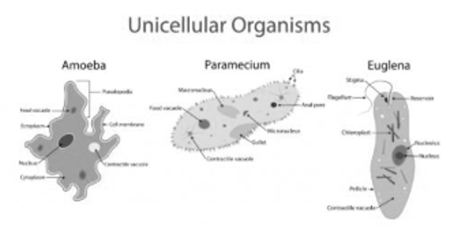
Fig 1: Shows pictures of unicellular organisms, Their Internal structure generally consists of a nucleus, plasma membrane, cilia, mitochondria, vesicles, and other cell organelles. Though they are free living they are dependent on other sources for their food. They eat up bacteria and other organic matter.
Archaea: These are another set of prokaryotes which significantly differ from bacteria. They occur in harsh climates like salt water and hot springs. however, they are also found in soils and oceans.
"Species are groups of potentially interbreeding natural populations which reproduce in isolation from other such groups."
This definition brings into view that the individuals of a single species are able to interbreed but stay isolated reproductively from each other.
Speciation cannot occur without reproductive isolation. In order to diverge from ancestral population, populations need to be divided for many generations of off spring and become new and independent species. However, if the population does not divide, itself either physically through some sort of barrier, or reproduce by their behaviour or follow other mechanisms like prezygotic or postzygotic isolation mechanisms, then the species will continue to be one species and will not divide itself into any other form and become its own distinct species. This isolation is central to the biological species concept.
Morphological Species
Morphology accounts for the individual looks an organism has. It is their physical features and anatomical parts. When Carlous Linnaeus first came up with his binomial nomenclature taxonomy, all individuals were grouped according to phenotype or morphology. Therefore, we must remember that the first concept of the term "species" was based on the morphology or phenotype. The morphological species concept does not take account into aspects like genetics and the DNA. Linnaeus was unaware about the chromosomes and their differences in evolution that actually make some individuals that look similar or a part of different species.
The morphological species concept definitely has its own limitations. First, it does not show any differences between species that are actually produced by different evolution and are not really related closely. It also does not group individuals of the same species that would happen to be somewhat morphologically different like in colour or size. It is much more accurate to use behaviour and molecular evidence to determine what is the same species and what is not.
Lineage Species
A lineage has a simple meaning of a branch on a family tree. The phylogenetic trees of that grow in many directions are actually groups of related species branch off where new lineages are created from speciation of a common ancestor. Subsequently some of them fail to exist over time while others become extinct others thrive and continue to live on. This lineage study of species becomes very important for scientists who are interested to study the history of life on earth and evolution.
On observing scientists can determine when the species diverged and evolved compared to their, factors like common ancestor the similarities and differences of different lineages are also taken into consideration. The asexually reproducing species can be studied under the concept of lineage species. Species that reproduce asexually is not much considered, Since biological species concept is dependent upon reproductive isolation of sexually reproducing species. The lineage species concept does not have that restraint and therefore can be used to explain simpler species that do not need a partner to reproduce.
Strains
In biology, a strain is a genetic variant, a subtype or a culture that exists within a biological species. Specific Genetic isolation are characterized by inherently artificial concept that Strains are often seen as in this aspect. This is most easily observed in microbiology in a petri dish where strains are derived from a single cell colony and are typically grown in isolation by the physical constraints of a petri dish. Strains are also commonly stated if fields of virology, botany, and also with rodents which are extensively used in experimental studies. By mutation or genetic swapping new viral strains can be created when two or more viruses infect the same cell. These phenomena are known respectively as antigenic drift and antigenic shift. Metagenomic methods are used to maximize resolution within species, this method is also used to differentiate Microbial strains by using their genetic makeup. This has become a valuable tool to analyse the microbiome. In biotechnology, a number of metabolic pathways have been established using microbial strains which has been constructed been for treating a variety of applications. Production of biofuel has been going on, Historically, a major effort of metabolic research. Strains of E. coli are extensively used for this application. Simple protein expression is obtained through E. coli. These strains, such as BL21, are modified genetically to minimize protease activity, hence large-scale protein production which is efficient can be enabled.
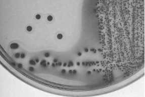
Fig 2: shows a strain of E. coli grown on an agar plate, some strains of E. coli can have a variety of virulence factors including adhesins, hemolysins, invasins, and toxins. The serotype O157:H7 (O refers to somatic antigen; H refers to flagellar antigen) is responsible for food poisoning causing haemorrhagic diarrhoea and haemolytic uremic syndrome (kidney failure).
Yeasts are extensively used for Industrial fermentation are the most common subjects of eukaryotic genetic modification.
E. coli is most common prokaryotic species used in strain engineering. Scientists have succeeded in developing a new strain with viable minimal genomes from which. These minimal strains provide a near guarantee that experiments on genes outside the minimal framework will not be affected by non-essential pathways.
Classification helps to describe the bacterial species based on diversity and grouping these organisms based on their similarities. Microorganisms can be classified on the basis of metabolism of cell and structure of the, or on differences in cell components such as fatty acids, DNA, fatty acids, antigens, antigens and quinones.
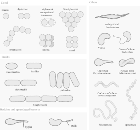
Fig 3: Bacterial Morphology: Basic morphological differences between bacteria. The most often found forms and their associations.
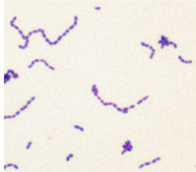
Fig 4: shows a species of bacteria Streptococcus mutants stained as gram positive rods
Microscopy: The observation of an enlarged picture of a minute organism is possible through a microscope, an instrument which provides a bigger or amplified image of an object that is not visible with the naked eye.
Aside from the common microscopy, there are a few special types of microscopy that include:
The word "microscopy" comes from Greek roots: mikros, small + skopeo, to view = to view small (objects).
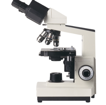
Fig 5: Shows the picture of a commonly used Compound microscope.
The magnifying power of a microscope is an expression of how many times the object being examined appears to be enlarged. The ratio does not have dimensions. It is usually expressed in the form 10× (for an image to be magnified 10-fold), The resolution of a microscope is expressed in linear units usually micrometres (μm). The resolution of a microscope is the measure of the smallest detail of the object that can be observed. Resolution is expressed in linear units, usually micrometres(μm).
The light microscope or the optical microscope is the most popular type of microscope, in this case glass lenses are used to form the image. Optical microscopes can be simple, or compound consisting of a single lens, or compound, consisting of several optical components or lenses in a line. The commonly used hand magnifying glass can magnify images to about 3 to 20×. Single-lensed simple microscopes can magnify images up to 300×—and are capable of revealing minute bacteria—while compound microscopes can magnify up to 2,000×. A simple microscope can show a resolution of objects below 1 micrometre (μm; one millionth of a metre); on the other hand, a compound microscope can show images with a resolution as low as about 0.2 μm.
Protozoa are the important source of food for the macroinvertebrates. As the protozoa are components of the micro and meiofauna, Thus, in order to help bacteria and algae to move successfully to higher tropic levels the ecological role of protozoa is very important. As predators, they prey upon filamentous algae, unicellular or bacteria, and micro fungi. Both herbivorous and consumers are included in the Protozoan species in the decomposer link of the food chain. They also control bacteria populations and biomass to some extent.
For all ecosystems, the fundamental source of energy is radiant energy from the sun. The energy of sunlight is used by the ecosystem’s autotrophic organism, or self-sustaining, organisms. Consisting largely of green vegetation, these organisms are capable of preparing their own food through photosynthesis—i.e., they can use the energy of sunlight to convert carbon dioxide and water into simple, energy-rich carbohydrates (glucose). The energy stored in the simple energy rich carbohydrates are used to produce more complex organic compounds like lipids, starches, proteins that control most of the life processes. The autotrophic segment of the ecosystem is commonly referred to as the producer level.
If an object or a surface needs to be cleared of all forms of organisms or germs then it has to undergo a process called, Sterilization itis the process of killing all forms organisms from the desired surface or object. In microbial terms it applies to killing microorganisms of all kinds like fungi, bacteria, yeast moulds and even viruses from desired surfaces.
Sterilization is a process of destruction of all forms of living microorganisms from a substance.
Articles that come in direct contact with humans and animals are subjected to sterilization.
These articles can include drugs, nutraceuticals, surgical equipment, food, etc. Sterilization is mainly a process carried to preserve the substance for a longer period of time and not allowing it to decay.
Secondly, a substance that is not sterile may cause infections when consumed or administered as it may contain microbes.
So, sterilization is a very essential process. The microbes are invisible to naked eye, and some bacteria even have a protective sheath on their surface making them resistant to even sterilization.
For this effective sterilization techniques are designed and studied in microbiology.
The Methods of Sterilization include
1. Heat methods.
2. Chemical sterilization.
Heat method of sterilization: This is the most common method of sterilization. The heat is used kills the microbes present in the substance. The extent of sterilization is affected by factors like, the temperature of the heat and duration of heating.
In heat sterilization process, as the temperature of heat goes up the time span required for sterilization decreases, therefore for a given temperature the longer the exposure to heat the better is the sterilization. Further, the sterilization time increases with a decrease in temperature and vice-versa. The most important factor is to maintain minimum sterilization time or minimum contact time for the heat to be in touch with microbes or bacteria and thereby kill them.
The heat method of sterilization is again of two types based on the type of heat used.
A) Moist heat methods
B) Dry heat methods
Moist heat method of sterilization: In moist heat sterilization heat is applied in the form of steam or just boiling to a high temperature. This method includes techniques like
Boiling is a most preferred method for metallic devices like needles, surgical scissors, scalpels, etc. Here substances are boiled to a constant temperature to sterilize them.
Pasteurization was discovered by Louis Pasteur; it is the process of heating the milk to a temperature of 6o degrees or 72 degrees 3 to four times. Here alternative heating and cooling kill all the microbes and melds without boiling the milk.
Using Steam (autoclaving): Here the substances are subjected to sterilization in an instrument called autoclave a steam sterilization equipment. The process is carried out at a temperature of 121degrees for 20 mins or 115 degrees for 60 min at 15psi pressure.
The saturated steam is formed at boiling temperature of water, at about100 degrees.
This steam produced in the Autoclave condenses on the material to be sterilised and relieves the latent heat repeatedly to convert back into the water.
Further, there is no possibility of any microbes being alive as the saturated steam under pressure percolates through all the narrow spaces killing all the microbes thereby making the sterilization very efficient and effective.
It is the most common method used for pharmaceutical drugs as it is powerful enough even to kill bacterial spores, bacterial spores are very inert and form a rigid cover that protects the cell wall during harsh conditions, by steam sterilization even the spores are killed.
Dry heat methods: This method includes the substances that are subjected to dry heat like
Flaming is the process of exposing metallic devices like scalpels, needle, scissors to flame for few minutes. The fire directly burns the microbes and other dust on the instrument.
Incineration is done especially lops used for inoculation, these inoculating loops are used in microbial cultures. The metallic end of the loop is heated to red hot on the flame. This exposure kills all the germs present on the loop.
Hot air oven is suitable for dry material like powders, metal devices, glassware’s, etc
Radiation method involves exposing the packed materials to radiation for sterilization. They are like UV rays that are kept at the entrance of door to prevent entry of live microbes that enter through air.
Media composition
Media composition is a very important aspect in microbiology as it provides all the medium in which the organism can grow its also called as in vitro or lab propagation or growth of animal cells under controlled condition. Media composition is selective and requires a procedure required for an essential step to culturing of cells celled or media selection for appropriate growth of cells. The media contain all the essential nutrients to make it available for the cells to growth freely. animal cells can use to both type of media can artificial media and natural media.
Artificial media-
This media is also called a defined media it contains all fully defined media components. the media should be maintained to physiological pH at 7. also, should be sterilized before use.
Natural media-
This media includes contents like body fluid like serum, plasma, amniotic fluid, etc. before usage the toxicity of the media has to be checked. Some selective media may also contain tissue extracts like liver, spleen etc.
Serum-free media- This media is free of serum and the main advantage of this media, is it can control the growth of organism by changes in the composition of media, for e.g., cell differentiation.
Basic Components of a media include-
Ingredients | Quantity (100 mL) |
Peptone | 0.5 g |
Yeast Extract | 0.2 g |
Sodium Chloride | 0.5 g |
Agar | 1.5 g |
pH | 7 |
Example of a table showing, the ingredients and the quantity, that appears on a media container.
The selected strain Streptococcus mutants used in the present study was maintained on nutrient agar media, Composition of nutrient agar media.
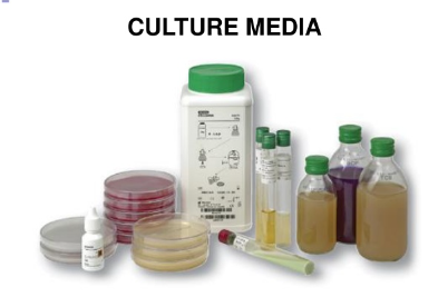
Fig: 6: picture shows the various culture media used for microbial growth.
In autecological studies, the growth of bacteria (or other microorganisms) in batch culture can be modelled with four different phases: lag phase (A), log phase or exponential phase (B), stationary phase (C), and death phase (D).
During lag phase, bacteria adapt themselves to conditions for growth. It is the period where the individual bacteria are maturing and still are not yet ready to divide. During the lag phase of the bacterial growth cycle, synthesis of RNA, enzymes and other molecules occurs. The cells are usually dormant during this stage, in the lag phase changes that occur in the cell are very minute, because the cells do not immediately reproduce in a new medium. This period of little to no cell division is called the lag phase and can last for 1 hour to several days.
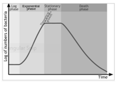
Fig 7: Shows the growth kinetics in bacteria
References: