Unit - 7
Macromolecular Analysis
Traditionally, science has taken a reductionist approach, science has been involved in dissecting biological systems into their constituent parts and studying them in detail and in isolation. The entire scientific careers have been devoted to studying only one protein or gene in order to understand its structure and function. The reductionist approach limits the insights in the human body although scientists have made progress using this method. As a result, limited success has been achieved by the efforts to treat many complex diseases.
Reductionism, by its nature, cannot be detailed or compared with the complexity of biological systems, the properties of these biological systems cannot be explained or predicted by studying their individual components. Subsequently these biological systems that are studied by scientists, now understand that the individual components of biological systems such as molecular pathways never work alone—they function in highly structured and integrated biological networks.
The changing properties and dynamics of these biological systems occur due to health and disease. Thus, a more holistic approach is required to gain a true understanding of health and disease, including the integrated analysis at broadly desperate levels, from molecular to organismal, from genetic to environmental. When scientists unravel this complexity, decipher these dynamic network interactions, and develop predictive models, then an understanding to life opens up.
All systems are working in synergy to analyse the multiple scientific developments and to advance systems biology. Firstly, new tools and technologies in today’s world is enabling analysis of complex networks of the changing dynamics that define healthy and disease states. Secondly, the enormous amount of data for storing and distributing has become easier. Last but not least, an expanded ecosystem of scientists is breaking down research silos and working together across disciplines.
- Protein Structure
Proteins are very important molecules and building blocks that are essential for all living organisms. Proteins are the largest unit of cells when analyse by dry weight, each protein is eventually devoted to a specific function and Proteins are involved in virtually all cell functions, with tasks that range from locomotion and cell signalling and general cellular support. In total, there are seven types of proteins the structure of a protein may be fibrous or globular depending on its specific role (every protein is specialized). Globular proteins are generally soluble, compact, and spherical in shape. Fibrous proteins are typically elongated and insoluble. Globular and fibrous proteins may exhibit one or more types of protein structures.
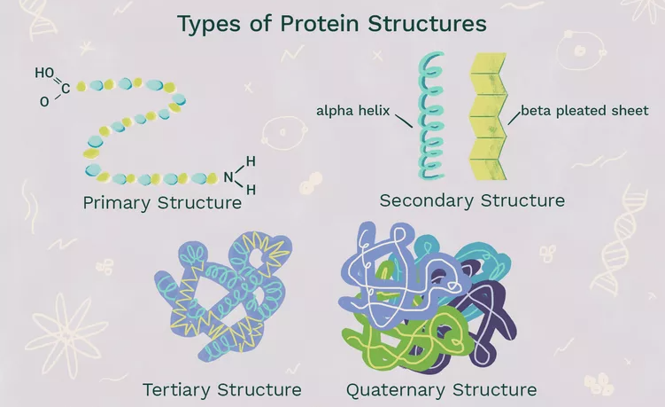
There are four structural proteins: namely primary, secondary, tertiary, and quaternary. These levels are determined by the complexity that lies in the polypeptide chain this nature differentiates them from each other, the shape and function of a protein. The primary level is the most basic and rudimentary while the quaternary level demonstrates complex bonding.
The countless nature of a protein determines its specific structure and function. A single protein molecule may contain one or more of these protein structure level. For example, Collagen, has a super-coiled helical shape that is strong, long, stringy, and rope-like collagen is very important for providing support. Haemoglobin, that is present in the blood, on the other hand, is a globular protein that is folded and compact. Its spherical shape is helpful for manoeuvring through blood vessels.
- Protein functions
Proteins play an important role in many crucial functions and biological processes. They are very versatile and have many different functions in the body, as listed below:
- They act as catalysts
- Transport other molecules
- They store other molecules
- Provide mechanical support
- Provide immune protection
- Generate movement
- Transmit nerve impulses
- Control cell growth and differentiation
The changes in the structure of a protein occurs when minute changes occur in their function or structure, the effect of the changes in protein is seen to such an extent to which the structure of proteins has an impact on their effect of changes and function in them. Any change to a protein at any structural level, including slight changes in the folding and shape of the protein, may render it non-functional.
- In particular, Laddertron Lang in 1952 first proposed that there was a hierarchy of protein structure with four levels: primary, secondary, tertiary, and quaternary. This hierarchy of protein is much easier as this structural classification is very convenient and anyone could start with this concept to teach the basic protein structure and this is probably familiar with this hierarchy as this structural classification is the easy and dependable. To revise: each level of the hierarchy is 'held together' by characteristic interactions and forces
- Higher levels of structure in the hierarchy are composed of the structural entities (also called elements) of the lower levels
- Primary protein structure
Proteins are made up of a long chain of amino acids. In the human body they are around 20amino acids that are commonly seen with a limited number of amino acid monomers. The proteins can be arranged in enormous number of ways to the function and alter the three-dimensional structure of the protein. The primary structure consists of the simple sequencing of the protein.
- Secondary protein structure
The secondary protein structure depends on the local interactions that occurs between parts of a protein chain, this can affect the folding and three-dimensional shape of the protein. There are two main things that can alter the secondary structure:
α-helix: sN-H groups in the backbone form a hydrogen bond with the C=O group of the amino acid 4 residues earlier in the helix.
β-pleated sheet: In this structure the N-H groups that lies in the backbone of one strand form hydrogen bonds with C=O groups in the backbone of a fully extended strand next to it.
Proteins can be linked with several functional groups such as alcohols, carboxamides, carboxylic acids, thioesters, thiols, and other basic groups linked to each protein. These functional groups also affect the folding of the proteins and, thereby, its function in the body.
- Tertiary structure
The tertiary structure of proteins, the proteins three-dimensional shape is taken into consideration, after the secondary interactions. These include the influence of non-polar, polar, acidic, and basic R groups that exist on the protein.
- Quaternary protein
The quaternary protein structure in this case refers to the arrangement and orientation of subunits in proteins that possess multi-subunits. This is only relevant for proteins with multiple polypeptide chains.
According to the sequence of amino acids in a polymer the Proteins subsequently fold up into specific shapes, and the protein function is directly related to the resulting 3D structure.
Proteins are also known to interact with each other or other macromolecules present in the body to create complex structures. In these assemblies, proteins can develop certain functions that would not have been possible as an individual protein, such as carrying out DNA replication and the transmission of cell signals.
The nature of proteins is also showing a lot of variation. For example, some are quite rigid, whereas others are somewhat flexible. These characteristics in proteins also makes them capable for the function of the protein. For example, more rigid proteins can play a role in the structure of the connective tissues or cytoskeleton. On the other hand, those with some flexibility may act as hinges, springs, or levers to assist in the function of other proteins.

Fig 1: The hierarchy of Proteins showing four different levels
Enzymes are proteins that facilitate and speed up biochemical reactions, which is why they are often referred to as catalysts. Notable enzymes include lactase and pepsin, proteins that are familiar for their roles in digestive medical conditions and specialty diets. Lactose intolerance is caused by a lactase deficiency, an enzyme that breaks down the sugar lactose found in milk. Pepsin is a digestive enzyme that works in the stomach to break down proteins in food—a shortage of this enzyme leads to indigestion.
Other examples of digestive enzymes are those present in saliva: salivary amylase, salivary kallikrein, and lingual lipase all perform important biological functions. Salivary amylase is the primary enzyme found in saliva and it breaks down starch into sugar.
Enzymes regulate almost all the numerous and complex biochemical reactions that occurs in plants, animals, and microorganisms. These catalytic proteins are efficient and specific—which means, they are specific and they accelerate the rate of only one kind of chemical reaction of one type of specific compound, and their analysis is far more efficient manner than human-made catalysts. The proteins are controlled by inhibitors and activators that initiate or block reactions. All cells contain enzymes, which usually are different in number and composition, and also depends on the cell type; for example, an average mammalian cell, is approximately one one-billionth (10−9) the size of a drop of water and generally contains about 3,000 enzymes.

Fig 2: Shows the various steps involved during the process of enzyme action
Transporters
A transport protein (that is referred in many terms as a transmembrane pump, transporter, acid transport protein, escort protein, cation transport protein, or anion transport protein) is a protein whose main purpose is to move other materials within an organism. Transport proteins are vital for the growth and life of all living organisms.
Transport Protein Definition
Transport proteins are proteins that mainly function to transport substances across biological membranes. The occurrence of proteins is found to be within the membrane itself, where they form a channel, or a carrying mechanism, wherein they allow their substrate to pass from one side to the other end.
The substances include sodium and potassium ions also sugars like glucose, messenger molecules, proteins and many more are transported by these proteins.
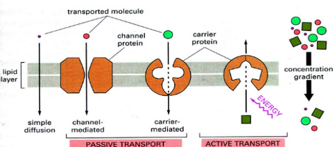
Transport proteins generally perform transport that is divided into two ways of transport: “facilitated diffusion,” where a transport protein creates an opening for a substance to diffuse down its concentration gradient; and “active transport,” where the cell expends energy in order to move a substance against its concentration gradient.
Function of Transport Protein
As we know certain important molecules, such as DNA, must be present inside the cell always; however other molecules such as ions, sugars, and proteins, can move in and out of the cell to function properly.
Each transport protein is designated to transport a specific substance when required. For example, some channel proteins, open only when they receive the correct signal, allowing the substances they transport to flow on demand. Active transporters, likewise, can often be “turned on and off” by messenger molecules.
Nerve impulses and cellular metabolism are possible by moving substances across membranes by transport proteins.
These proteins are so very important as the transport proteins, for example, the sodium-potassium gradient that allows our nerves to fire would not exist.
Types of Transport Proteins
- Channels/Pores
As their name suggests, “channel” or “pore” proteins help in opening holes in the membrane of a cell.
These proteins are characterized by being open at the same time for both the intracellular and extracellular activities. In contrast, to carrier proteins which are only open to the inside or outside of a cell at any specific time.
Channels or pores are typically designed in such a way that only one specific substance can pass through.
For example, voltage-gated ion channels often use charged amino acids, that are present at specific distances, to attract their desired ion while all the others are repelled. The desired ion can then flow through the channel while other substances cannot.
A good example of transport proteins are the Voltage-gated ion channels are that act specifically. They are often seen in neurons, voltage-gated ion channels open in response to changes in a membrane’s electrochemical potential.
When closed, the voltage-gated channel does not allow any ions to pass through the cell membrane. But when open, it allows huge quantities of ions to pass through very quickly, allowing the cell to change its membrane potential rapidly and fire a nerve impulse.
- Carrier Proteins
Carrier proteins are transport proteins that are only open to one side of the membrane at a particular time.
As they transport substances against the concentration gradient. They are often designed this way. If the membrane is open simultaneously to both sides it might allow these substances to simply flow back along their concentration gradient, putting an end to the carrier protein work.
To achieve their work, carrier proteins change their shapes and utilise the energy.
When an example of the sodium-potassium pump, is considered , it uses the energy of ATP to change its shape and opens itself to the extracellular solution where it was initially open to the intracellular solution, resulting of which allows it to collect ions inside the cell and release them outside of it, and then vice versa.
Other sources of energy are used by other carrier proteins that may include, existing concentration gradients, this energy is used to achieve “secondary active transport.” The protein does not use the ATP directly but the transport is made possible through the cell expending energy, but the protein itself does not use ATP directly.
A unique feature of the carrier proteins is that they often use the energy present in one substance that wants to undergo a change in its shape therefore it moves down its concentration gradient. The change in shape of the substance allows it to transport a substance that “doesn’t want” to move at the same time.
The concentration gradient of sodium that is used by the sodium-glucose transport protein is a very good example – which was originally created by the sodium-potassium pump – where the glucose was moved against its concentration gradient.
We discuss the sodium-potassium pump and the sodium-glucose transport protein in detail below.

- Receptors
Receptor Definition
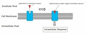
A receptor is a protein which binds to a specific molecule. The molecule to which a receptor binds is known as the ligand. A ligand may be any molecule, that ranges from from inorganic minerals to hormones to organism -created proteins, neurotransmitters. The ligand binds to the ligand-binding site on the receptor protein. The receptor undergoes a conformational change when this binding happens, the protein function is slightly altered by the change in shape of the receptor. Following this a number of things can happen. During such conformational change in the receptor, the receptor can become an enzyme and actively separate or combine certain molecules.
The changes in the receptor can also cause a series of changes in related proteins, eventually transmitting some sort of message to the cell. This message could be a sensory signal or it could be metabolic regulation message. Binding affinity is the capacity of the receptor to hold onto the ligand. Eventually the receptor will release the ligand once the attraction dies down and the receptor will change to its original shape, and the message or signal will end. The series of processes that take place depends on the strength of the affinity between the receptor and ligand.
Apart from the receptor other molecules can also attach to the ligand-binding site on a receptor. These molecules are called agonist molecules and if they show similar effect of the natural ligand. Many drugs, which may be prescribed or illegal taken, are synthetic agonists to molecules one such example is endorphins, these drugs create feelings of satisfaction. It is noticed that more than the natural ligand these molecules often have a stronger affinity for the receptor. This explains why the tolerance to certain drugs or painkillers can stay for a longer time as the agonist will remain attached to the receptor for a longer duration, therefore a much higher dose is required to block this activity
Sometimes the receptor does not undergo any conformational change when it binds to a ligand because other molecules act like antagonists, these molecules block the ligand binding site on the receptor and does not allow the receptor to undergo any change. Eventually blocking a signal entirely. Some doctors use these drugs (receptor antagonists) to deprive people of alcohol dependency and heroin, these receptor antagonists make the drug no longer a delight. Certain proteins present in snake venom that imitate platelet binding protein are achieved by the antagonists. Therefore, the receptors which would normally connect platelets and prevent bleeding are therefore disabled. This can lead to internal bleeding and death. Effective medicines are being created by Pharmaceutical companies in both agonists and antagonists for their potential.
Types of Receptors
In the mammalian body there are literally thousands of different types of receptors. While they are endless to list out, receptors do fall into some very broad categories of function. Many are used in “cellular signalling”, cellular signalling is a vast complex system of signals and responses that is mediated entirely by receptors and the ligands they receive. These include receptor proteins embedded in the cellular membrane which activate other sequences upon receiving a ligand, and the receptors found in the immune system which are structured to find intruding proteins and molecules. Below is the general model for cell signalling, which can take many different forms.

- Proteins as structural elements
Structural proteins provide rigidity and stiffness to otherwise-fluid biological components. Most structural proteins are fibrous protein; for example, collagen and elastin are critical components of connective tissue such as cartilage, and Keratin some of the examples where keratin are mainly found are hair, nails, feathers, hooves, and some animal shells which are usually hard and filamentous structures ,globular proteins actin and tubulin are globular and soluble as monomers, which polymerize to form stiff and long fibres that make up the cytoskeleton, and help the cell to maintain its shape and size.
Myosin, dynein and kinesin are motor proteins that serve as structural and functional proteins capable of generating mechanical forces. These proteins play a very crucial for cellular motility of single celled organisms and the sperm of many multicellular organisms which reproduce sexually. These proteins help in intracellular transport and also generate the forces exerted by contracting muscles.
Proteins, such as collagen, are important for the structure of tissues and serve as the scaffolding of the body.

Fig 3: Collagen when viewed under the microscope looks like a long fibre that are woven together to provide extra strength, collagen almost accounts for about a quarter of the protein present in the body.
Collagen is the most important structural protein found in cells skin and bones. Structural proteins are also used to build structural components of the body, such as bones and cartilage.
In cells the structural protein provides internal structure to the cell and also help in cell movement. Structural proteins are important in larger cells and are involved in cellular movement.
Collagen when viewed under the microscope looks like a long fibre that are woven together to provide extra strength, collagen almost accounts for about a quarter of the protein present in the body.
Collagen provides elasticity of skin in the absence of collagen the skin appears wrinkled. The main function of collagen is to support tissues in the body and to provide structure for specific types of cells. The body uses collagen in blood vessels, ligaments and tendons.
All proteins are built by smaller molecules to form large molecules, these small molecules are called amino acid, when these amino acids join together in various combinations, they form proteins with different properties. Other types of proteins include hormones, enzymes and carrier molecules.
Other types of structural proteins include keratins that form protective covering for the skin, hair, nails, claws, beaks, feathers etc. Actins and myosins, are found in muscle tissue and the silks and insect fibres. The hides of vertebrates and the tendons are places where the collagen is also present, they form connective ligaments in the body and provide extra support to various parts of the skin.
References:
1.Proteins Structure and Function: David Whitford
2. Protein Stability and Folding- Bret.A. Shirley
3. How proteins work- Mike Williamson.